3D liver models for drug testing

Regulatory agencies, such as the Food and Drug Administration (FDA), are moving towards reducing animal testing and encouraging the use of New Alternative Methodologies (NAMs) in pharmaceutical research.
NAMs are non-clinical tests that can provide information on chemical hazards and risk assessment without using animals, and they include in silico, in chemico, in vitro, and ex vivo approaches.
3D cell cultures are promising NAMs for extracting reliable conclusions in drug testing and liver-related pathologies. Let’s see it in detail!
.
Why move towards 3D co-culture liver models?
Liver cell culture models have advantages over animal models regarding cost, ethical considerations, and efficiency.
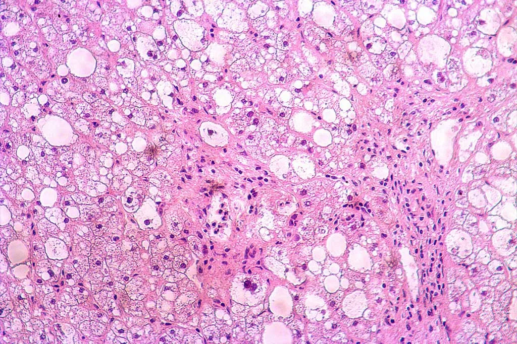
The liver is a complex hepatocyte-formed organ, representing approximately 70% of its total cell population. Non-parenchymal cells (NPCs) represent the remaining 30% and include liver endothelial cells/liver sinusoidal endothelial cells (LECs/LSECs), Kupffer cells (KCs), and hepatic stellate cells (SCs).
2D cell cultures are easy to establish, but they fail to recapitulate the intricate physiology of the human liver. Drug metabolic enzymes, such as cytochrome P450, are less stable under 2D conditions, and hepatocyte metabolic activity is lower.
On the other hand, 3D monocultures, although better at recreating the liver structure, do not reflect the crosstalk between parenchymal and non-parenchymal cells.
The presence of NPCs is crucial for sustaining the functionality of liver cells in an in vitro setting, increasing the expression of CYP and Phase II isoforms compared to monotypic culture.
Accurate liver models must replicate the structure and function of the liver in vivo as much as possible to become in vitro tools that closely predict drug outcomes in human health. No liver models are precise without including NPCs and facilitating cell-cell and cell-extracellular matrix (ECM) contact in 3D.
.
Examples of 3D co-culture liver models
3D co-culture models can be classified according to the contact between the cells conforming to the model:
1- Direct 3D co-culture. These models include two or more cell types in close contact, allowing communication through cell-cell adhesion, paracrine secretion of soluble cytokines, and cell-ECM adhesion.
Some examples are self-aggregating multicellular spheroids, 3D liver organoids, and co-cultures of liver cells in 3D scaffolds of natural or synthetic materials.
2- Indirect 3D Co-culture. Two or more liver cell types are cultured in a 3D environment with a physical separation system that does not allow physical contact. This separation system is usually a Transwell chamber that allows paracrine signaling interactions between cells through soluble cytokines. Transwells avoid unnecessary contact between cells and facilitate separation for single-cell analysis.
3- Mixed methods. Mixed methods combine direct and indirect cultures, where some cells exhibit cell-cell contact, and the remaining populations are physically separated.
These methods include 3D bioprinting co-culture, which allows for the spatial arrangement of cells, and microfluidic multicellular liver chips, which reproduce physiological oxygen and nutrient gradients.
.
Empower your 3D cell co-culture liver models with BeCytes
At BeCytes, we are experts in providing primary hepatic cells to create accurate models in vitro that mimic liver physiology.
Our extensive portfolio offers human primary hepatocytes, fresh or cryopreserved, and human non-parenchymal cells, including Kupffer Cells, liver endothelial cells/liver sinusoidal endothelial cells, and stellate cells.
All our cells are produced under rigorous QC standards and supplied with:
-
- Demographic and clinical donor profile
- Viability and morphology assessment
- Extensive characterization depending on cell type
- Specific certificate of analysis
- Culture and maintenance protocols
- Defined media for each cell type
It has never been easier to generate 3D co-culture liver models. BeCytes Biotechnologies provides you with all the cellular components.
Contact us with any questions or for personalized assistance at mailto:becytesinfo@bioivt.com.
References
Ma Y, Hu L, Tang J, Guo W, Feng Y, Liu Y, Tang F. Three-Dimensional Cell Co-Culture Liver Models and Their Applications in Pharmaceutical Research. Int J Mol Sci. 2023 Mar 26;24(7):6248. doi: 10.3390/ijms24076248
Stresser DM, Kopec AK, Hewitt P, Hardwick RN, Van Vleet TR, Mahalingaiah PKS, O’Connell D, Jenkins GJ, David R, Graham J, Lee D, Ekert J, Fullerton A, Villenave R, Bajaj P, Gosset JR, Ralston SL, Guha M, Amador-Arjona A, Khan K, Agarwal S, Hasselgren C, Wang X, Adams K, Kaushik G, Raczynski A, Homan KA. Towards in vitro models for reducing or replacing the use of animals in drug testing. Nat Biomed Eng. 2023 Dec 27. doi: 10.1038/s41551-023-01154-7.


