5 reasons to include Stellate Cells in your fibrotic liver model

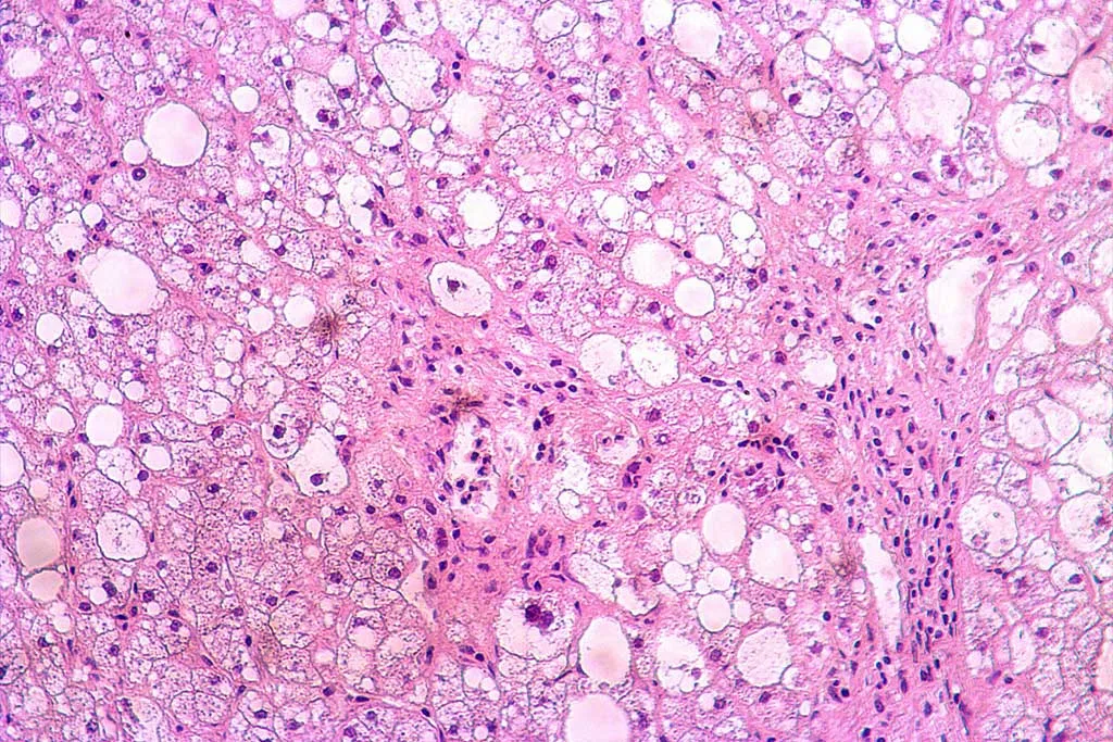
Human hepatic stellate cells (HSCs) are liver-specific mesenchymal cells located in the space of Disse, between the endothelial cells of the sinusoid and the hepatocytes. Although HSCs represent only 5–10% of all resident cells within the liver, they perform numerous critical functions in healthy and injured liver.
During their inactive state, HSCs contain numerous lipid droplets of vitamin A, constituting the body’s largest reservoir. In response to injury, HSCs transdifferentiate and change phenotype. This process is key in fibrotic liver disease, such as metabolic dysfunction-associated steatotic liver disease (MASLD), formerly known as non-alcoholic fatty liver disease (NAFLD), and its progression to metabolic dysfunction-associated steatohepatitis (MASH), previously known as nonalcoholic steatohepatitis (NASH).
Here are 5 reasons to consider including stellate hepatic cells in your fibrotic in vitro model. Keep reading until the end for an extra one!
.
1. Role in fibrosis development
HSCs show a quiescent and an active state. In response to liver injury, HSCs activate and transdifferentiate from an adipogenic to a myofibroblast phenotype with loss of lipid droplets and expression of contractile fibers. HSCs, through diverse signaling processes, enhance the local accumulation of extracellular matrix capable of forming a scar in areas of liver injury. This activation is a crucial factor leading to MASLD progression, so including HSCs in fibrotic models is necessary to understand the fibrogenesis process.
.
2. Fat metabolism and storage
While hepatocytes are the main cells involved in fat accumulation in MASLD, HSCs also play a role in lipid metabolism and storage, particularly regarding vitamin A. Understanding the dynamics of fat metabolism in stellate cells can provide insights into the overall lipid homeostasis within the liver in MASLD.
.
3. Inflammatory and immune response
MASLD and especially its progression to MASH involve significant inflammatory and immune responses. Stellate cells produce various cytokines upon activation and can influence immune cell recruitment. Their presence is crucial to illuminating the inflammatory pathways involved in MASLD progression.
.
4. Therapeutic targets
Due to their central role in fibrosis, stellate cells are key targets for therapeutic interventions in MASLD /MASH. Prevention of stellate cell activation or induction of their deactivation is crucial for developing treatments to prevent fibrosis in MASH.
.
5. Biomarkers identification
Models that include stellate cells can aid in identifying biomarkers of disease progression from MASLD to MASH and fibrosis. Biomarkers are essential for early diagnosis, disease progress monitoring, and assessing therapeutic interventions’ response.
.
6. Easy access to HSCs
Easy access to hepatic stellate cells is the final reason you need to add them to your fibrotic model.
At BeCytes, we are experts in providing primary liver cells to assist researchers in creating advanced cellular models that mimic the intricate microenvironment of the human body.
Our extensive portfolio offers diverse hepatic cell types, from non-parenchymal cells to MASLD hepatocytes. Check the available lots by selecting the cell type and adjusting the parameters. If you have any doubt, don’t hesitate to contact us at becytesinfo@bioivt.com.
Don’t hesitate to contact us!
.
References
Kamm DR, McCommis KS. Hepatic stellate cells in physiology and pathology. J Physiol. 2022;600(8):1825-37.
Wang M, Li L, Xu Y, Du J, Ling C. Roles of hepatic stellate cells in NAFLD: From the perspective of inflammation and fibrosis. Front Pharmacol. 2022;13:958428..
.


