Isolation and culture of human primary hepatocytes
")
Isolation and culture of human primary hepatocytes
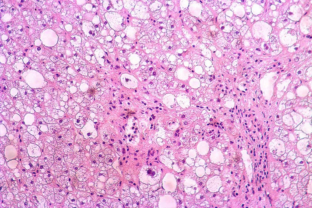
Primary human hepatocytes are considered the gold standard for liver cell studies due to their ability to closely mimic the physiological functions of the liver. Unlike cell lines such as HepG2, primary hepatocytes maintain the full range of metabolic activities, including proper cytochrome P450 enzyme expression, bile acid synthesis, and drug transport mechanisms when properly cultured.
However, isolation and difficult access are significant challenges when working with primary hepatocytes. Obtaining a consistent supply of high-quality hepatocytes used to be a problem, limiting their widespread use in research and drug development. Nevertheless, improvements in isolation protocols and cell providers like BeCytes Biotechnologies have solved this problem.
.
How to isolate human primary hepatocytes
Gone are the days when mechanical methods were the only option to isolate liver cells. Filtering through a cheesecloth or shaking with glass beads damaged hepatocytes and led to recovery yields between 5 and 10 %.
In 1962, the enzymatic perfusion method was introduced using collagenase and hyaluronidase perfused through the liver portal vein in rats. This perfusion method has been refined and improved, leading to a two-step procedure for liver cell isolation, which is the gold standard, especially in the case of large liver samples.
- Step 1: Pre-perfusion with a calcium-free buffer. The liver or liver tissue fragment is perfused with a calcium-free buffer, such as Hank’s Balanced Salt Solution (HBSS) with EGTA, to disrupt desmosomes that form tight junctions between cells. EGTA also helps to remove any residual blood.
- Step 2: Enzymatic digestion with collagenase in a buffer containing Ca2+. Several collagenases have been tested for hepatocyte isolation. Protocols can include sequential perfusions with buffers containing a mixture of collagenase class I and II, collagenase D, or collagenase IV.
After the digestion, the liver is carefully removed to obtain the cell suspension that is then filtered through a 3-layer nylon mesh to separate undigested tissue remnants.
Two-step perfusion protocols have reported a yield of hepatocytes with cell viability of 77 ± 10% (Lee et al. 2013). Optimized protocols have recently been published to isolate either normal or non-alcoholic steatohepatitis livers (Liu et al. 2023).
Mechanical dissociation is combined with enzymatic digestion in resected healthy human liver tissue which is unsuitable for perfusion, especially for smaller fragments. Green et al. propose a protocol where tissue is diced and undergoes a two-step digestion with EDTA and collagenase. This method has been reported to obtain >65% viable hepatocytes despite using smaller liver tissue pieces.
.
BeCytes isolates human primary human liver cells for you
At BeCytes, we are experts in isolating and characterizing primary human liver cells. We take care of cell isolation and characterization so you can focus on your research. After many years of scientific research, we have developed a proprietary isolation protocol that allows us to obtain human primary hepatocytes with a granted post-thawing viability superior to 85%.
We have mastered the protocols to grant you access to human primary hepatocytes, including MASLD, and non-parenchymal cells, sourced from meticulously characterized donor tissues. Rigorous testing ensures they meet the highest viability, purity, and functional activity standards.
All our primary liver cells are produced under QC standards and supplied with:
- Demographic and clinical donor profile
- Viability and morphology assessments
- Characterization by using specific expression markers
- Specific Certificate of Analysis (CoA) with culture and maintenance protocols
- Specific cell culture media
Our CoA now includes organoid generation capability! Besides having a section for spheroid formation, the CoA includes a new section describing whether a specific batch of human primary hepatic cells can generate liver organoids. This information is available for both primary hepatocytes and non-parenchymal cells.Check our portfolio out!
References
Green CJ, Charlton CA, Wang LM, Silva M, Morten KJ, Hodson L. The isolation of primary hepatocytes from human tissue: optimising the use of small non-encapsulated liver resection surplus. Cell Tissue Bank. 2017 Dec;18(4):597-604. doi: 10.1007/s10561-017-9641-6.
Kaur I, Vasudevan A, Rawal P, Tripathi DM, Ramakrishna S, Kaur S, Sarin SK. Primary Hepatocyte Isolation and Cultures: Technical Aspects, Challenges and Advancements. Bioengineering (Basel). 2023 Jan 18;10(2):131. doi: 10.3390/bioengineering10020131.
Lee SM, Schelcher C, Demmel M, Hauner M, Thasler WE. Isolation of human hepatocytes by a two-step collagenase perfusion procedure. J Vis Exp. 2013 Sep 3;(79):50615. doi: 10.3791/50615.
Liu X, Lam K, Zhao H, Sakane S, Kim HY, Eguileor A, Diggle K, Wu S, Gontijo Weber RC, Soroosh P, Hosseini M, Mekeel K, Brenner DA, Kisseleva T. Isolation of primary human liver cells from normal and nonalcoholic steatohepatitis livers. STAR Protoc. 2023 Sep 15;4(3):102391. doi: 10.1016/j.xpro.2023.102391.
.


