How are HepG2 cells different from primary hepatocytes?

How are HepG2 cells different from primary hepatocytes?
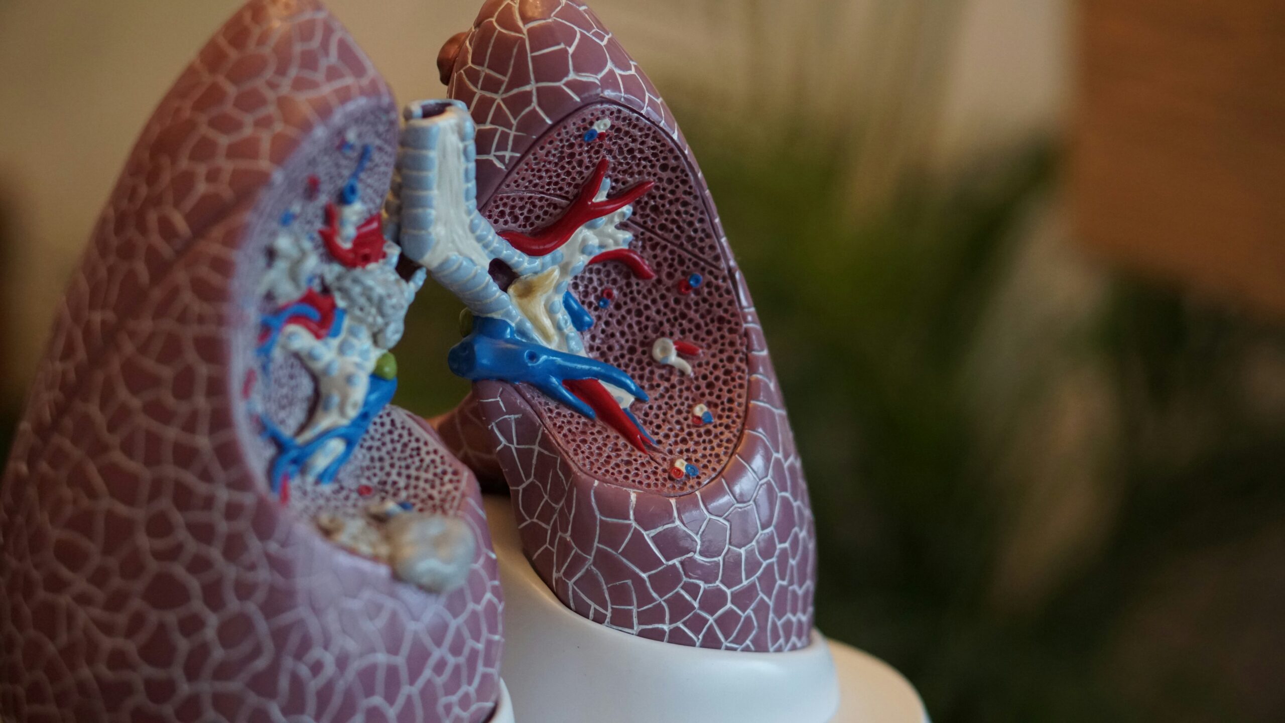
Cellular models can make all the difference in biomedical research. In particular, the liver’s complex functions require models that accurately reflect its broad-spectrum functionalities.
When it comes to choosing between widely used options like HepG2 cells and primary hepatocytes, the differences between these models can significantly influence the direction and outcomes of your research. So, let’s delve into the key differences between these two cellular models.
.
HepG2 cells are a convenient but limited model
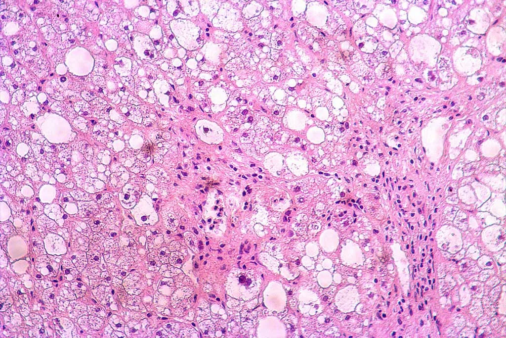
HepG2 cells are a human liver cancer cell line extensively used in research due to their easy accessibility and maintenance. These cells have certain advantages, including their ability to proliferate indefinitely, which makes them an attractive option for long-term studies. Moreover, they are relatively easy to transfect, making them a useful tool in genetic studies. However, the limitations of HepG2 cells become critical when studying complex liver functions or drug metabolism.
Since HepG2 cells are derived from hepatocellular carcinoma, they exhibit altered metabolic pathways that do not fully represent the behaviour of normal liver cells. For example, the expression levels of key drug-metabolizing enzymes (i.e., cytochrome P450) are significantly lower in HepG2 cells compared to primary hepatocytes. Along with their altered responses to hormonal and inflammatory signals, this makes them less suitable for studying drug metabolism, liver toxicity, and metabolic diseases. Moreover, HepG2 cells show deficiencies in bile acid synthesis and transport, have reduced expression of liver transporters essential for drug absorption and excretion, and their cancerous origin leads to genomic instability, rendering their experimental results less reliable than those from primary hepatocytes.
Moreover, HepG2 cells fall short of replicating the liver’s complex, multicellular environment. Interactions between various cell types, such as Kupffer cells and endothelial cells, are crucial for accurately modelling liver function, a context that HepG2 cells are unable to fully recreate.
.
Try the gold standard for liver research with primary hepatocytes
In contrast, primary hepatocytes, which are isolated directly from liver tissue, offer a more physiologically relevant model for liver function studies. These cells retain the metabolic activities and cellular structures of their tissue of origin, providing a more accurate representation of liver function.
Primary hepatocytes are often considered the gold standard for drug metabolism, toxicity, or liver disease studies.
One of the primary advantages of using primary hepatocytes is their ability to maintain the expression of key liver-specific enzymes and proteins, including the full spectrum of cytochrome P450 enzymes. This allows for more accurate studies on drug metabolism and interactions, providing data that is more likely to translate into human clinical outcomes. Additionally, primary hepatocytes exhibit normal liver cell morphology and function, including albumin secretion and urea synthesis, which are critical for studying liver physiology and pathology.
Another significant benefit of primary hepatocytes is their ability to respond to stimuli in a way that closely mimics in vivo conditions. This includes their response to inflammatory cytokines, hormones, and other signalling molecules, which is essential for studies on liver inflammation, fibrosis, and other diseases.
BeCytes’ primary human hepatocytes, sourced from meticulously characterized donor tissues, provide exceptional reliability and reproducibility for your experiments. Rigorous testing ensures they meet the highest standards in viability, purity, and functional activity. Our hepatocytes deliver the accuracy and clinical relevance you need for drug metabolism, toxicology, or liver disease research.
Reach out today and take your liver research further with BeCytes.
References
Guo L, Dial S, Shi L, Branham W, Liu J, Fang JL, et al. Similarities and differences in the expression of drug-metabolizing enzymes between human hepatic cell lines and primary human hepatocytes. Drug Metab Dispos. 2011 Mar;39(3):528–38.
LeCluyse EL, Witek RP, Andersen ME, Powers MJ. Organotypic liver culture models: meeting current challenges in toxicity testing. Crit Rev Toxicol. 2012 Jul;42(6):501–48.
Donato MT, Gallego-Ferrer G, Tolosa L. In Vitro Models for Studying Chronic Drug-Induced Liver Injury. Int J Mol Sci. 2022 Sep 28;23(19):11428.
Rodríguez-Antona C, Donato MT, Boobis A, Edwards RJ, Watts PS, Castell JV, et al. Cytochrome P450 expression in human hepatocytes and hepatoma cell lines: molecular mechanisms that determine lower expression in cultured cells. Xenobiotica. 2002 Jun;32(6):505–20.
.


