Predict Drug-Induced Liver Injury (DILI) with Primary Human Hepatocyte Spheroids
 with Primary Human Hepatocyte Spheroids")
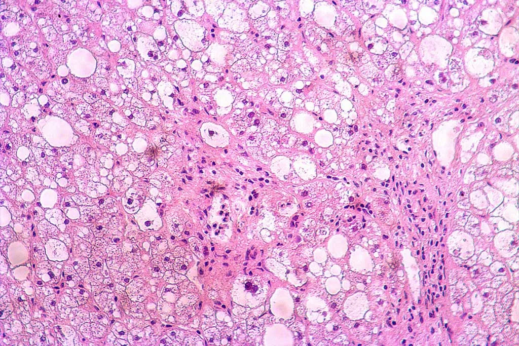
Drug-induced liver injury (DILI) has fatal consequences, both personally and economically. Its health impact ranges from mild liver enzyme elevation to severe liver failure. In the drug development process, it is a leading cause of drug withdrawal, resulting in significant costs for pharmaceutical companies in terms of direct financial impacts and lost opportunities.
Accurate preclinical models are essential to predict DILI during drug discovery and development. Regulatory agencies encourage reducing animal testing and using New Alternative Methodologies (NAMs), and 3D liver models are the perfect fit to understand the complex mechanisms underlying DILI.
Stern, Wang, and Sadrieh include Primary Human Hepatocyte (PHH) spheroids as a key microphysiological model to predict DILI, highlighting their experimental relevance. Let’s explore it!
.
Advantages of Primary Human Hepatocyte Spheroids
Primary Human Hepatocytes are the gold standard for studying liver biology and are widely used in preclinical studies. Their self-assembly by gravity-induced aggregation generates liver spheroids that present multiple advantages:
-
- They sustain hepatic cellular phenotypes and viability in extended culture for 4-5 weeks.
- PHH spheroids present relevant 3D architecture maintaining cell-cell interactions.
- They are very representative of the native liver compared with other liver models in 2D and 3D culture formats.
- PHH spheroids are cost-effective and adaptable to a higher throughput workflow.
To maximize their mimicking properties, liver spheroids can combine PHH with non-parenchymal cells (NPCs), such as Kupffer Cells, Liver Sinusoidal Endothelial Cells (LSEC), and Stellate Cells (SC).
13 % of acute liver failures generated by DILI have an unknown cause and are considered idiosyncratic. Immune mechanisms mediate these cases. Kupffer Cells, specialized macrophages resident in the liver, are often overlooked as components in preclinical models to predict DILI. Spheroids combining PHH, Kupffer cells, and stellate cells can be used to model chronic exposure with an increased sensitivity of hepatocytes to detect clinically relevant concentrations of hepatotoxins.
Adding LSECs to PHH spheroids increases CYP activity and urea secretion compared to spheroids with hepatocytes alone. The presence of endothelial cells improves the functional maturity of hepatocytes and promotes polarization and 3D architectural arrangement.
.
3D primary human hepatocyte spheroids vs 2D cultures to predict DILI
3D PHH spheroids show increased sensitivity of in vitro liver toxicity assays. Li, Parikh, and Zuo tested 100 selected DILI and control compounds and compared their predictive power in 3D PHH spheroids to 2D monolayer cultures of PHHs.
In many cases, 2D monolayer cultures failed to detect liver toxicity of the DILI compounds. Compared to them, 3D PHH spheroids were 2-3 times more sensitive in detecting hepatotoxicity in severe, high, and low-concern DILI compounds with repeated dosing.
Their study also elucidates that a ratio of 2:1 of primary human hepatocytes and Kupffer Cells can recapitulate inflammatory responses in a 3D culture system that is more physiologically relevant.
The addition of LSECs also results in better predictive responses. Proctor et al. demonstrated that spheroids combining PHH, KCs, and LSECs enhance sensitivity to identify hepatotoxic drugs compared to a 2D hepatocyte monoculture after screening a panel of 110 marketed drugs.
.
BeCytes aims to be your partner in creating reliable preclinical models
Reliable preclinical models are indispensable in drug discovery and development as they provide critical insights into a drug’s toxicity, efficacy, and safety before human trials. They help minimize risks to human subjects and guide the design of clinical trials, thereby increasing the likelihood of regulatory approval and market success, reducing time and cost while maximizing therapeutic potential.
BeCytes aims to be a fundamental partner for creating reliable preclinical models. We are experts in providing human and animal hepatic cells. We can provide primary hepatocytes together with NPCs from the same donor.
Our human primary hepatocytes are 3D-certified to generate spheroids. Check out our inventory to find the perfect lot for your 3D cell culture. Our tested positive lots for 3D culture are labeled as 3D/Spheroid Qualified.
.
Contact us with any questions or for personalized assistance at becytesinfo@bioivt.com.
References
Ardalani H, Sengupta S, Harms V, Vickerman V, Thomson JA, Murphy WL. 3-D culture and endothelial cells improve maturity of human pluripotent stem cell-derived hepatocytes. Acta Biomater. 2019 Sep 1;95:371-381. doi: 10.1016/j.actbio.2019.07.047.
Bell CC, Hendriks DF, Moro SM, Ellis E, Walsh J, Renblom A, Fredriksson Puigvert L, Dankers AC, Jacobs F, Snoeys J, Sison-Young RL, Jenkins RE, Nordling Å, Mkrtchian S, Park BK, Kitteringham NR, Goldring CE, Lauschke VM, Ingelman-Sundberg M. Characterization of primary human hepatocyte spheroids as a model system for drug-induced liver injury, liver function and disease. Sci Rep. 2016 May 4;6:25187. doi: 10.1038/srep25187
Li F, Cao L, Parikh S, Zuo R. Three-dimensional spheroids With Primary Human Liver Cells and Differential Roles of Kupffer Cells in Drug-Induced Liver Injury. J Pharm Sci. 2020 Jun;109(6):1912-1923. doi: 10.1016/j.xphs.2020.02.021
Proctor WR, Foster AJ, Vogt J, Summers C, Middleton B, Pilling MA, Shienson D, Kijanska M, Ströbel S, Kelm JM, Morgan P, Messner S, Williams D. Utility of spherical human liver microtissues for prediction of clinical drug-induced liver injury. Arch Toxicol. 2017 Aug;91(8):2849-2863. doi: 10.1007/s00204-017-2002-1.
Stern S, Wang H, Sadrieh N. Microphysiological Models for Mechanistic-Based Prediction of Idiosyncratic DILI. Cells. 2023 May 25;12(11):1476. doi: 10.3390/cells12111476.


