In vitro skin models as an alternative to cosmetic animal testing

In vitro skin models as an alternative to cosmetic animal testing
Animal testing for cosmetic products has been banned in the European Union since 2013. This includes a prohibition on the sale of cosmetics tested on animals outside the EU. Other countries have followed this prohibition or implemented restrictions, such as Brazil, Canada or Australia.
The United States has made strides toward banning animal testing for cosmetics, though it is not yet fully implemented nationwide. The Humane Cosmetics Act has been reintroduced and continues to gain support.
The global outlines define increasing international efforts to adopt and promote the use of New Alternative Methods for cosmetic testing.
.
The skin: a complex organ that requires complete models
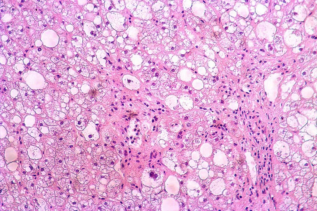
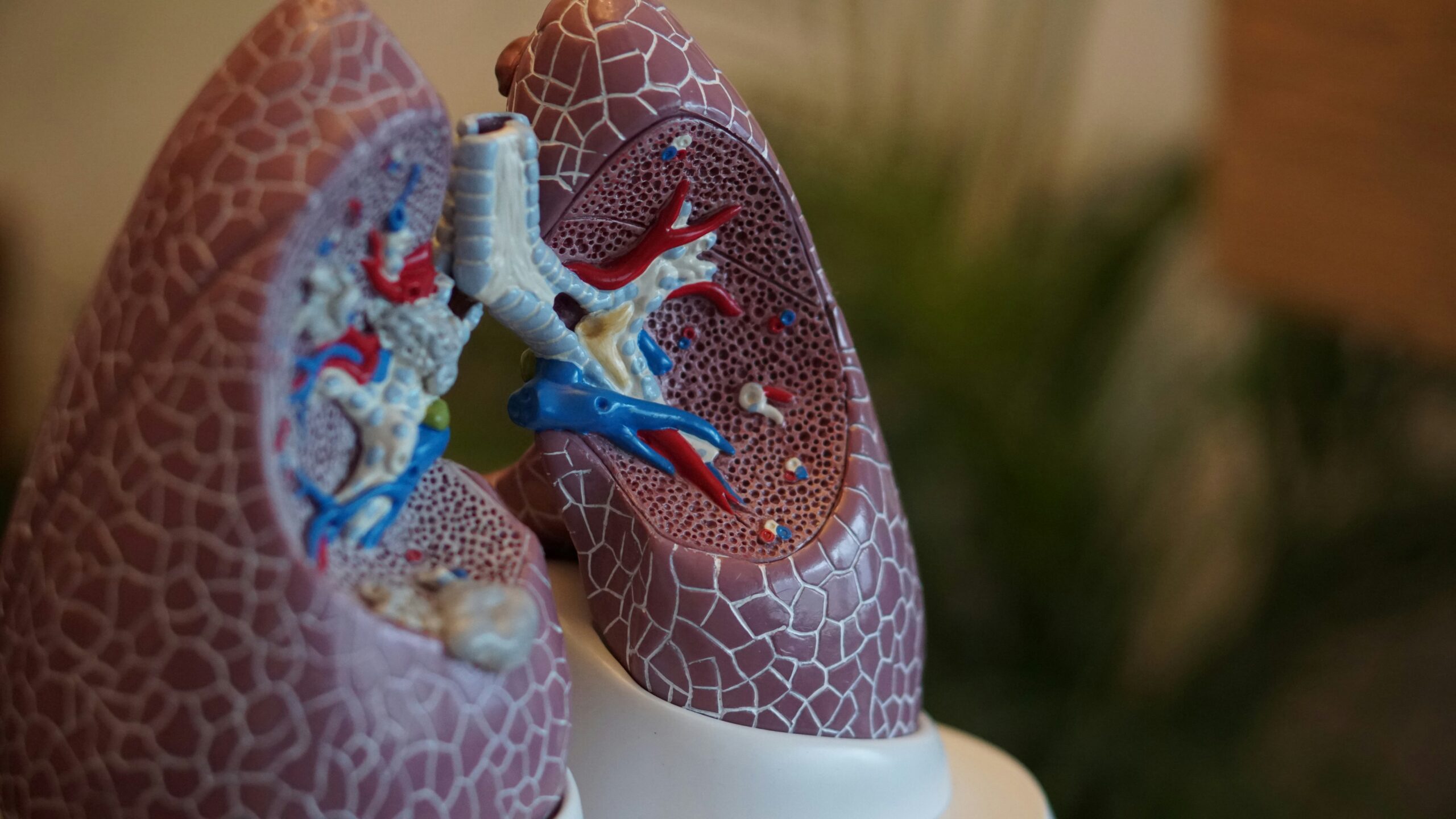
The skin is the largest organ of the human body, consisting of three main layers: the epidermis, the dermis, and the hypodermis. The interconnection of the layers and their cell types (keratinocytes, melanocytes, fibroblasts, adipose cells…) ensures the skin’s health and functionality.
This complex, interconnected system needs to be carefully reproduced in vitro to provide viable alternative models to replace cosmetic animal testing.
2D in vitro cell culture models have been the alternative to animal use in performing conventional cosmetic screenings. Despite its popularity, 2D models fail to recapitulate skin complexity because they consist of cells seeded as a monolayer. Nutrient distribution is not well represented in the monolayer as it occurs in the native thick tissue. Besides, it does not favor cell-cell and cell-extracellular matrix interaction. A great example of this model is the reconstructed human epidermis (RHE) which comprises only the epidermal layer of the skin and was the first commercially available reproduction of human skin.
In the 80s, a bi-layered human skin model (HSM) containing keratinocytes seeded on top of a collagen gel comprised of fibroblasts was developed. It was the first organ-mimicking system introduced, but despite the complexity increase, this kind of model still lacks vascularization and certain cell types.
.
3D in vitro systems for skin modeling
3D models have the potential to recapitulate skin complexity offering tissue equivalents with multiple cell types, accurate structure, and a vascularization system. The development of organoids and organ-on-a-chip technologies has opened a wide range of possibilities for the dermatological and skin-care industries.
.
Organoids
Stem cells are the most common source to derive skin organoids. The skin contains a variety of adult stem cells (ASCs), including epidermal, hair follicle, mesenchymal stem cells, melanocytes …
Besides ASCs, pluripotent stem cells, including embryonic stem cells (ESCs) and induced ones (iPSCs) are also a source of skin organoids.
Organoids require a matrix-rich 3D environment to self-organize into a tridimensional structure. This can be accomplished using commercial Matrigel, natural or synthetic hydrogels, as well as various scaffold materials. Furthermore, 3D printing technology can be utilized to replicate the solid structures of the stratum corneum or basement membrane, ensuring accurate spatial organization of cells.
Despite their vast potential, the use of skin organoids has been focused on the study of early human skin development and the modeling of skin diseases. This model still faces some drawbacks, such as the inability to culture in air–liquid interface. It hinders accurate epidermal differentiation and makes it a challenge to use organoids for topical drug/cosmetic treatments.
.
Organ-on-a-chip
Skin-on-a-chip systems can be developed using two different methods:
-
- Direct introduction of a skin fragment in the microfluidic system. Diverse research has been published either using skin micro-biopsies or commercialized HSE models.
- In situ generation directly inside the chip. Cells self-organize inside the device, the channels being used to deliver nutrients and as compartments to hold the tissue. Diverse cell types and perfusion are introduced. Shear stress is produced in the system increasing cell viability and proliferation in contrast to static cultures.
Organ-on-chip complexity is leveled up by interconnecting multiple organ-like functional in vitro tissues to study further interactions.
.
BeCytes coordinates access to quality fresh skin biospecimens
At BeCytes, we are experts in coordinating access to fresh skin samples. We have created a platform capable of coordinating the donation of human tissues.
Do you need fresh tissue to establish organoids? Or to introduce it in your microfluidic chip?
Our extensive network promotes collaborations with national and international procurement centers, biobanks, and hospitals to provide researchers with various tissue samples and formats.
Send us an email to tissue_solutions@becytes.com and tell us about your research project. If there’s a match, we will find it!
.
References
Filaire E, Nachat-Kappes R, Laporte C, Harmand MF, Simon M, Poinsot C. Alternative in vitro models used in the main safety tests of cosmetic products and new challenges. Int J Cosmet Sci. 2022 Dec;44(6):604-613. doi: 10.1111/ics.12803.
Hong ZX, Zhu ST, Li H, Luo JZ, Yang Y, An Y, Wang X, Wang K. Bioengineered skin organoids: from development to applications. Mil Med Res. 2023 Aug 22;10(1):40. doi: 10.1186/s40779-023-00475-7
Rimal R, Muduli S, Desai P, Marquez AB, Möller M, Platzman I, Spatz J, Singh S. Vascularized 3D Human Skin Models in the Forefront of Dermatological Research. Adv Healthc Mater. 2024 Apr;13(9):e2303351. doi: 10.1002/adhm.202303351
Sun H, Zhang YX, Li YM. Generation of Skin Organoids: Potential Opportunities and Challenges. Front Cell Dev Biol. 2021 Nov 4;9:709824. doi: 10.3389/fcell.2021.709824


