How to culture liver organoids

How to culture liver organoids
Organoids are three-dimensional (3D) structures derived from stem cells, progenitors, or differentiated cells that self-organize to recapitulate aspects of the native tissue architecture and function, resembling mini-organs. Contrary to spheroids, organoids tend to organize themselves when placed in a matrix-rich 3D environment, requiring cell-matrix interactions and specific growth factors.
The increasing trend in culturing liver organoids is reflected in the number of papers published. A quick search in Pubmed indicates that 10 years ago, in 2014, just 21 papers were published containing the keyword “Liver Organoid”, in 2023 the number grew to 357.
Culture methods, including cell source, 3D matrices, and culture media, differ from one paper to another.
.
Cell source
Liver organoids can be derived from pluripotent stem cells (PSCs) or liver tissue, which includes primary liver hepatocytes and other cell types such as non-parenchymal cells.
Embryonic stem cells (ESCs) and induced pluripotent stem cells (iPSCs) present high pluripotency, plasticity, and infinite proliferation capacity. Nevertheless, ESCs present ethical concerns regarding their origin. iPSCs lead the race to become the leading in vitro model system, due to their unlimited proliferation, the ability to generate different cell types, and suitability for genome editing. But despite their potential, iPSCs reprogramming can generate epigenetic and genetic aberrations, including both chromosomic and aneuploidy alternations. Their high derivation and expansion cost is also a relevant drawback.
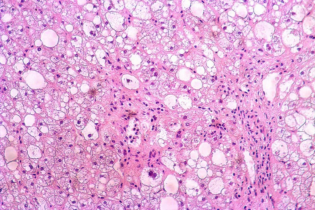
Primary hepatocytes constitute an ideal source for liver organoid establishment. Recent studies demonstrate their proliferation abilities and stemness. Primary hepatocytes offer a wide range of possibilities due to their ability to generate either hepatocyte or cholangiocyte organoids. Differentiation towards a particular lineage depends on cell selection, Lgr5+ ductal cells in the case of cholangiocyte organoids, and the differentiation cell culture media.
Besides their similarity to their origin organs, which provides a more comprehensive point of view, compared to iPSCs, primary tissue-derived organoids are more mature and have higher genome stability.
.
3D matrix
Matrigel, a commercial extracellular matrix (ECM), is the gold standard for organoid self-organization induction. Matrigel is secreted by Engelbreth-Holm-Swarm (EHS) mouse sarcoma cells, which makes its composition variable from lot to lot. It leads to biochemical and mechanical variability and a lack of reproducibility in organoid generation.
New alternatives comprising natural and synthetic hydrogels and decellularized ECM have emerged, but their adoption as a standardized 3D in organoid culture protocol is far from being adopted. Matrigel remains the choice for its accessibility, convenience, and versatility.
Culture media
Focusing on liver organoids derived from liver tissue, expansion medium, and differentiation medium are usually combined. The first allows cells to divide and grow into 3D structures and the second one is used to induce hepatocyte fate because these 3D structures present bipotentiallity both expressing bile duct and hepatocyte-lineage markers.
Hu et al. have greatly reviewed the functions and the main ingredients in both types of culture media used to culture tissue-derived liver organoids in a recently published paper.
BeCytes provides hepatic cells and tissues to establish liver organoids
Although much progress has been made, there is still a long road to standardizing protocols for liver organoid culture leading to reproducible 3D in vitro liver models.
BeCytes contributes to developing accurate 3D in vitro models for disease modeling and drug screening by providing human hepatic tissue and primary liver cells, including primary hepatocytes and non-parenchymal cells.
Along with a complete characterization, our cells are tested for their ability to form spheroids, indicated by a 3D/Spheorid column in our inventory. We are also working to provide a section describing whether a specific batch can generate liver organoids. It will be available soon.
Check our inventory! Find non-parenchymal cells to model liver microenvironment
.
References
Gunti S, Hoke ATK, Vu KP, London NR Jr. Organoid and Spheroid Tumor Models: Techniques and Applications. Cancers (Basel). 2021 Feb 19;13(4):874. doi: 10.3390/cancers13040874
Hu Y, Hu X, Luo J, Huang J, Sun Y, Li H, Qiao Y, Wu H, Li J, Zhou L, Zheng S. Liver organoid culture methods. Cell Biosci. 2023 Nov 1;13(1):197. doi: 10.1186/s13578-023-01136-x
Schneeberger K, Sánchez-Romero N, Ye S, van Steenbeek FG, Oosterhoff LA, Pla Palacin I, Chen C, van Wolferen ME, van Tienderen G, Lieshout R, Colemonts-Vroninks H, Schene I, Hoekstra R, Verstegen MMA, van der Laan LJW, Penning LC, Fuchs SA, Clevers H, De Kock J, Baptista PM, Spee B. Large-Scale Production of LGR5-Positive Bipotential Human Liver Stem Cells. Hepatology. 2020 Jul;72(1):257-270. doi: 10.1002/hep.31037..


